Peritoneal Dialysis (PD) catheter is a soft and flexible tube inserted into the peritoneum that allows the patient to perform peritoneal dialysis.
The PD catheters are made of polyurethane or silicone, and they may present 1 or 2 Dacron cuffs:
- 1st Cuff (know as Peritoneal Cuff): is placed into the muscle wall of abdomen;
- 2nd Cuff (known as Subcutaneous Cuff): is placed in the skin where the catheter comes out of the body.
These cuffs help the catheter to be in place, as the fibrous tissue will grow into them. They also act as a barrier preventing germs from going up to the catheter.

Before understanding the peritoneal dialysis process, it is essential to comprehend the concept of dialysis itself.
So, what is Dialysis?
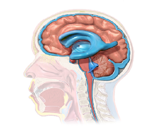
Dialysis is a blood cleansing process performed when patients are in terminal renal failure (end-stage); meaning that their renal function is less than 15% (see figure below). This process filters the patient’s blood of body waste, extra salt and water, and also helps to control the patient’s blood pressure.
 Note that there are 2 different types of dialysis:
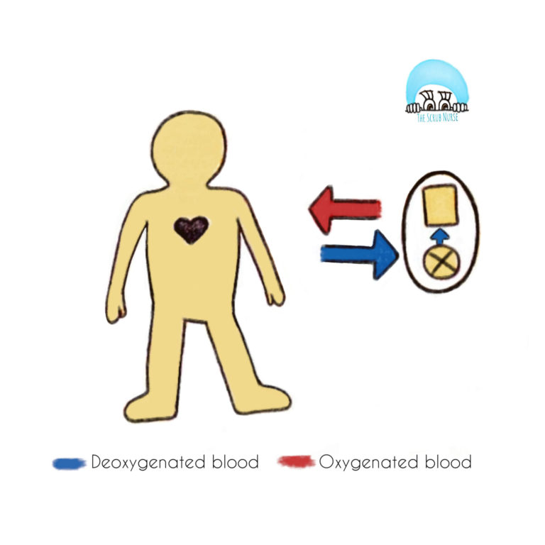
- Hemodialysis – when blood is pumped out of patient’s body into a dialysis machine and returns to the body after the filtration process is finished.
- Peritoneal dialysis (PD) – when the peritoneal cavity acts as a natural filter, by using a cleansing fluid that is slowly infiltrated in the peritoneum through a catheter. At the end, the fluid is drained along with body wastes to a drainage bag.

Now lets focus in the Peritoneal Dialysis:
Please attend the following concepts used in Peritoneal Dialysis:
- Dialysate – cleansing fluid;
- Dwell time – period of time required for the cleaning fluid to be within the peritoneum. Usually 4 to 6 hours;
- Exchange – the process of filling and draining the peritoneum.
How does Peritoneal Dialysis work?

- First, the dialysate flows into the abdomen and remains in the peritoneum for a prescribed dwell time;
- During that period, the dialysate enables the filtration process in which the body’s waste, chemicals and extra fluid are filtered from the peritoneum’s cappilaries.
- Finally, once the dwell time is finished, the solution inside of the peritoneum (that at this moment is composed by waste products drawn from the blood) is drained into the collection bag.
There are 2 different methods of peritoneal dialysis, depending on their exchange’s schedules:

Continuous Ambulatory Peritoneal Dialysis (CAPD):
- For every exchange, requires patient collaboration to connect a fresh bag of dialysis solution and to remove the drain solution bag;
- Does not require a machine;
- Uses gravity to fill and empty the abdomen;
- Every 4 to 6 hours of dwell time a new cycle of dialysis repeats (exchange);
- Requires 3 or 4 exchanges during the day and 1 long overnight (with a dwell time of 8 to 10 hours).
Continuous Cycling Peritoneal Dialysis (CCPD):
- Requires a machine to fill and empty the abdomen;
- Used during the night in which 3 to 5 exchanges occur;
- The last fill of the cleansing soltuion performed overnight remains in the abdomen during the whole day (long dwell time);
- Sometimes an additional exchange during the day may be required.
The surgical procedure
Now that the PD catheter and its role in patient’s life were explained, have a look in the surgical steps of its insertion (Open technique):
- Anaesthesia: GA
- Patient’s position: Supine
- Special equipments required: PD catheter and tunneling tool
- Infraumbilical midline incision is performed;
- Dissection of subcutaneous tissue, muscle and some adhesions, if present;
- Small peritoneum incision (1 to 2 cm) – to allow catheter to be placed;
- The catheter is pushed into the peritoneal cavity;
- The 1st cuff of the catheter is stitched in the preperitoneal space;
- The exit site of the catheter is choosen;
- A tunneling tool is used to create a tunnel under the skin until the exit point;
- The catheter is pulled through the tunnel;
- The Second cuff of the catheter is placed subcutaneously and stitched (2 cm from the exit site);
- The catheter is tested by filling the abdomen with 100ml of sterile saline and draining the fluid throught the catheter;
- The incision is closed.
References